Molecular epitopes of the ankyrin-spectrin interaction
Abstract
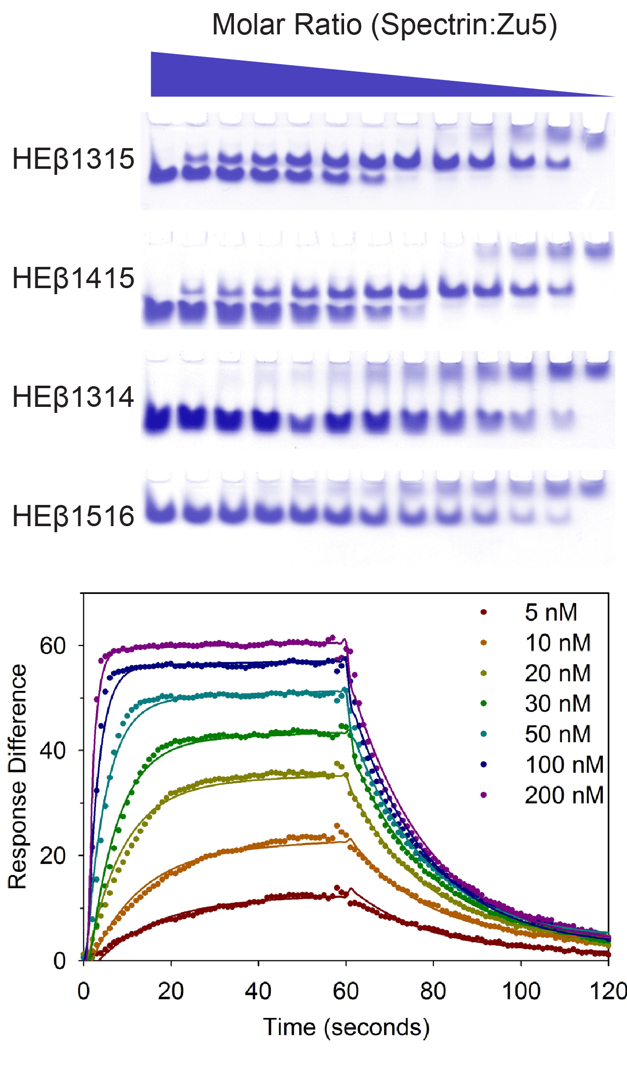 Isoforms of ankyrin and its binding partner spectrin are responsible for a number of interactions in a variety of human cells. Conflicting evidence, however, had identified two different, non-overlapping human erythroid ankyrin subdomains, Zu5 and 272, as the minimum binding region for beta-spectrin. Complementary studies on the ankyrin-binding domain of spectrin have been somewhat more conclusive yet have not presented binding in terms of well-phased, integral numbers of spectrin repeats. Thus, the objective of this study was to clearly define and characterize the minimal ankyrin-spectrin binding epitopes. Circular dichroism (CD) wavelength spectra of the aforementioned ankyrin subdomains show that these fragments are 30-60% unstructured. In contrast, human erythroid beta-spectrin repeats 13, 14, 15, and 16 (prepared in all combinations of two adjacent repeats) demonstrated proper folding and stability as determined by CD and tryptophan wavelength and heat denaturation scans. Native polyacrylamide gel electrophoresis (PAGE) gel shifts as well as affinity pull-down assays implicated Zu5 and beta-spectrin repeats 14-15 as the minimum binding epitopes. These results were confirmed by analytical ultracentrifugation to sedimentation equilibrium by which a 1:1 complex was obtained if and only if Zu5 was mixed with beta-spectrin constructs containing repeats 14 and 15 in tandem. Surface plasmon resonance yielded a K D of 15.2 nM for binding of beta-spectrin fragments to the ankyrin subdomain Zu5, accounting for all of the binding observed between the intact molecules. Collectively, these results show the 14th and 15th beta-spectrin repeats comprise the minimal, phased region of beta-spectrin, which binds ankyrin at the Zu5 subdomain with high affinity.